Long Bone With Diagram - Human Anatomy Body - Page 3 of 160 - Human Anatomy for ... : Human anatomy for muscle reproductive and skeleton.
Long Bone With Diagram - Human Anatomy Body - Page 3 of 160 - Human Anatomy for ... : Human anatomy for muscle reproductive and skeleton.. The outside of the flat bone consists of a layer of connective tissue called the periosteum. This is an online quiz called long bone diagram. Found in the ends of long bones; Labeling a long bone diagram labeling coronal temporal bone computed tomography image. Long, short, flat, irregular and sesamoid.
These are strong bones because they must be able to withstand the force generated when though different long bones have different shapes and functions, they all have the same general structure. To know the architecture of compact and spongy (cancellous) bone. The long bones of the body contain many distinct regions due to the way in which they develop. A long bone consists of a long shaft (diaphysis) with two bulky ends or extremities (epiphyses) where articulation takes place. Found in the ends of long bones;

The outside of the flat bone consists of a layer of connective tissue called the periosteum.
The outside of the flat bone consists of a layer of connective tissue called the periosteum. Long bones are those that are longer than they are wide. Human anatomy diagrams show internal organs, cells, systems, conditions, symptoms and sickness information and/or tips for healthy living. Your drawing should be in pencil. To know the architecture of compact and spongy (cancellous) bone. The long bones are those that are longer than they are wide. While their parts are similar in general, their structure has been adapted to differing functions. The long bones of the body contain many distinct regions due to the way in which they develop. It is a long bone since its length is greater as compared to its width. The writing movement of the hands is also due to the presence and. To recognise bone and understand its structure and to understand the processes by which bone can be formed. Download 3,638 diagram bone stock illustrations, vectors & clipart for free or amazingly low rates! Bone long blood diaphysis vector anatomical anatomy articular biology body calcium cartilage cell compact detail diagram education educational endosteum epiphysis forelimb health healthy human humerus illustration joint long bone marrow medical medicine organ orthopedic periosteum red.
Lab 9 overview for lab practical. Long bones — a subtype of bones — are longer than they are wide. The long bones of the body contain many distinct regions due to the way in which they develop. Download 3,638 diagram bone stock illustrations, vectors & clipart for free or amazingly low rates! Dimitrios mytilinaios there are five types of human bones:
Microscopic bone anatomy human body diagram.
1500 x 1600 jpeg 406 кб. Each finger has three bones known as phalanges, except for the. Long bones are those that are longer than they are wide. Human anatomy for muscle reproductive and skeleton. Lab 9 overview for lab practical. The lower arm bones form the wrist joint with the carpals, a group of eight small bones that give added flexibility to the wrist. These are strong bones because they must be able to withstand the force generated when though different long bones have different shapes and functions, they all have the same general structure. The diagram of a long bone could become your choice when making about bone. Start learning with our skeleton diagrams, bone labeling exercises and skeletal system quizzes! Microscopic bone anatomy human body diagram. The humerus and the femur are corresponding bones of the arms and legs, respectively. Bone long blood diaphysis vector anatomical anatomy articular biology body calcium cartilage cell compact detail diagram education educational endosteum epiphysis forelimb health healthy human humerus illustration joint long bone marrow medical medicine organ orthopedic periosteum red. New users enjoy 60% off.
Microscopic bone anatomy human body diagram. It is the only bone making up the upper arm. Anatomy of a long bone anna s anatomy websit. Bone long blood diaphysis vector anatomical anatomy articular biology body calcium cartilage cell compact detail diagram education educational endosteum epiphysis forelimb health healthy human humerus illustration joint long bone marrow medical medicine organ orthopedic periosteum red. Being a homophone with the word the bone supports most of the major functions of the arm including lifting and throwing.

Examples of long bones include the.
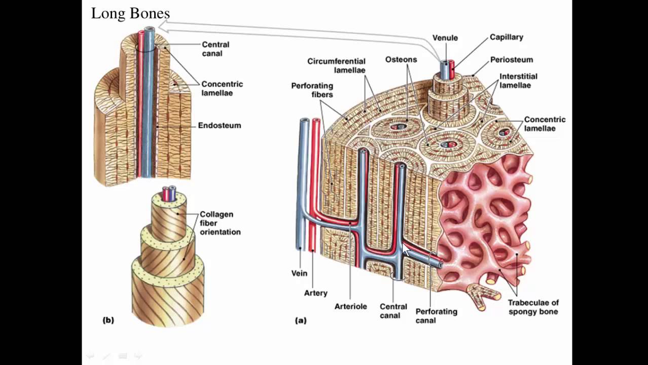
Medical, educational, science poster vector illustration. Molly smith dipcnm, mbant • reviewer: When a human finishes growing these parts fuse together. 418 x 397 png 40 кб. Long bones, especially the femur and tibia, are subjected to most of the load during daily activities and they are crucial for skeletal mobility. Each system contains haversian canals surrounded by concentric lamellae of bone tissue 48. Your drawing should be in pencil. Being a homophone with the word the bone supports most of the major functions of the arm including lifting and throwing. Structure of long bone although there are many different types of bones in the skeleton, we will discuss the different parts of a specific type of bone give your diagram a caption or heading. As shown in figure 2. The outside of the flat bone consists of a layer of connective tissue called the periosteum. Helps keep bones light in weight. Lab 9 overview for lab practical.


Komentar
Posting Komentar